Global Journal of Neurology and Neurosurgery received 111 citations as per Google Scholar report


Received: 01-Jul-2022, Manuscript No. GJNN-22-73063; Editor assigned: 04-Jul-2022, Pre QC No. GJNN-22-73063 (PQ); Reviewed: 18-Jul-2022, QC No. GJNN-22-73063; Revised: 25-Jul-2022, Manuscript No. GJNN-22-73063 (R); Published: 02-Aug-2022, DOI: 10.15651/2449-1942.22.10.009
The treatment of patients with brain tumors are typically multidisciplinary and involves a neurosurgeon, an oncologist, a radiologist, and a radiation therapy specialist. The patient's primary care physician is best able to coordinate the services of consultants, but the responsible neurosurgeon should be in charge of managing any postoperative issues or care that may arise. If a CNS tumour recurs, it might be necessary to transfer to a facility with qualified neurosurgical personnel.
The manner of treatment varies greatly depending on the location, type, and comorbidities of the tumour. A ventricular shunt can be installed, a tumour can be removed or debulked, and radioactive implants can be inserted.
Prehospital care focuses on the presenting symptom complex and is supportive. Treat seizures, for instance, as usual. Endotracheal intubation and, possibly, hyperventilation may be necessary for definitive airway control in cases of airway disturbance, breathing difficulty, signs of pronounced elevation in Intracranial Pressure (ICP), and notable impairment of consciousness.
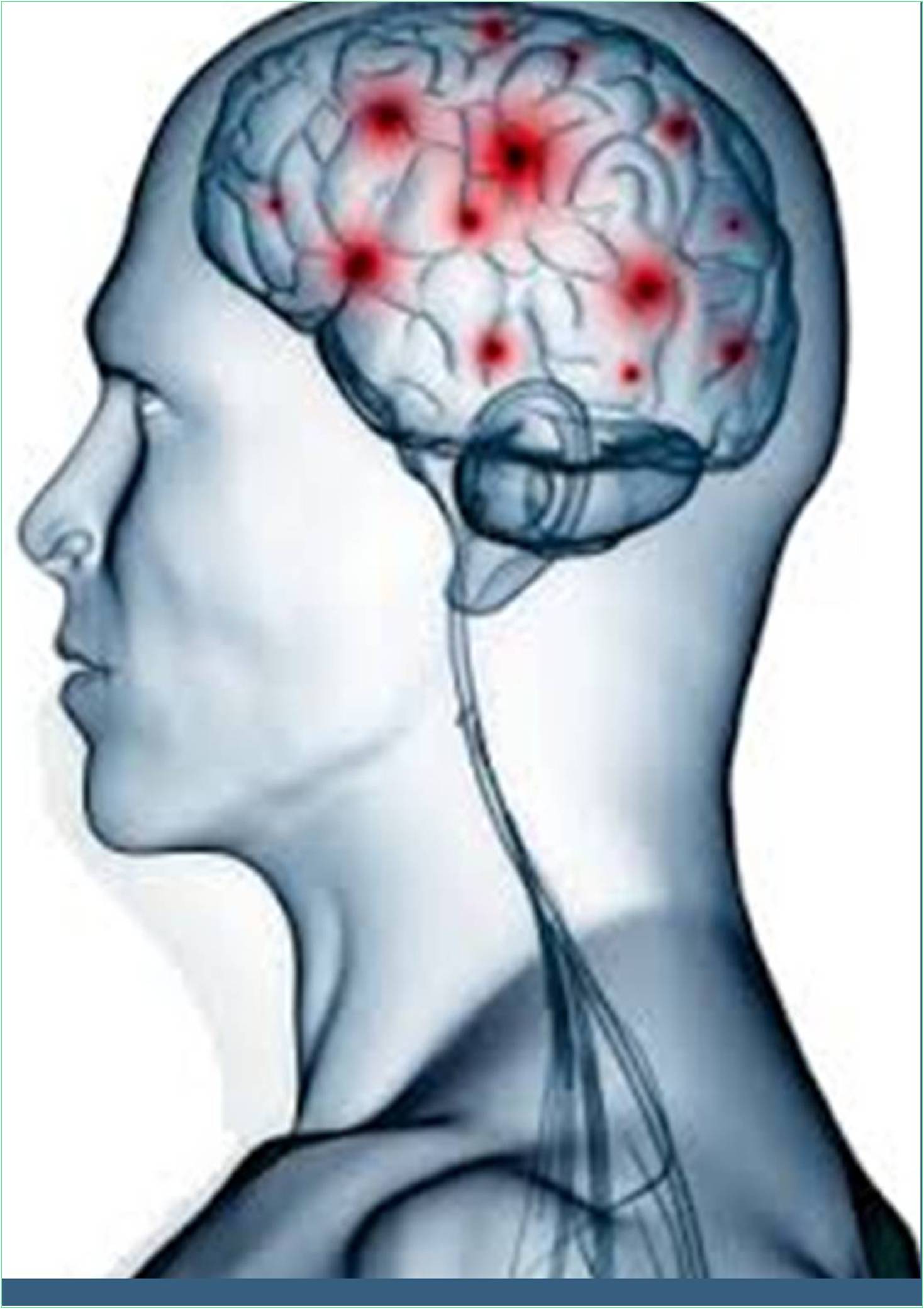
Treatment given to a patient with an intracerebral neoplasm in the Emergency Department (ED) depends on the tumor's type and the patient's overall health. The ED doctor is not qualified to make decisions about starting chemotherapy, radiation treatment, or surgical resection.
Patients with known brain tumours who complain of headaches or worsening symptoms are a common problem for ED doctors. The possibility of a tumour recurrence or worsening cerebral edoema is always present in this scenario. To rule out potentially fatal situations, such as hemorrhage or herniation, get a CT scan or MRI.
Corticosteroids may significantly lessen cerebral edemarelated signs and symptoms. Patients who receive steroid therapy may feel better within the first few hours.
The preferred agent is dexamethasone because it has negligible salt-retaining characteristics. The usual dose ranges from 4 to 24 mg per day. 10 mg IV or 10-24 mg IV is advised as the first dose for patients who exhibit signs of elevated Intracranial Pressure (ICP) or impaired consciousness. Dose-dependent side effects include proximal muscle weakness. After receiving definitive treatment, corticosteroids can frequently be reduced or stopped. The final steroid dosage should be as low as is required to manage the patient's neurological symptoms.
Consider using adjunctive medications for rapidsequence intubation in patients who show signs or symptoms of impending herniation or airway compromise. Lidocaine and drugs for rapid-onset neuromuscular blockade may be among them, along with measures to lessen fasciculations. Thiopental and other induction drugs may be used.
Consider mild hyperventilation after the airway has been successfully controlled.
With the appropriate consultant, go over mannitol usage. Mannitol may momentarily lower ICP, but its use is problematic due to worries about rebound increases in ICP.
Surgical Care
A qualified neurosurgeon must perform a tissue biopsy to provide a conclusive diagnosis. Options for neurosurgery include ventricular shunt placement with obstructive hydrocephalus as well as resection or debunking.
Complications
Acute symptoms in a patient with a brain tumour raise the possibility of an acute haemorrhage into the tumour, especially if the signs and symptoms resemble stroke symptoms. Lung cancer, melanoma, and choriocarcinoma are examples of brain tumours predisposed to bleeding.
Headaches, syncope, and changes in mental status are examples of paroxysmal symptoms brought on by lesions close to the third ventricle. Additionally, it's possible to experience vomiting, ataxia, memory loss, visual disturbances, or personality changes.
Temporary symptoms result from sporadic increases in ICP brought on by pressure brought on by a blockage of the cerebrospinal fluid outflow. A reported side effect of third ventricle outflow drainage obstruction is sudden death. The brain parenchyma will move in the direction of least resistance, which is caudally through the foramen magnum (posterior fossa tumours) or transtentorial apertures, if there are sudden increases in ICP.
Some pituitary tumours can cause acromegaly or galactorrhea because they are hormonally active. Pituitary apoplexy, an unusual side effect of pituitary adenomas, is characterised by bleeding into the tumour, which causes headache, blurred vision, oculomotor palsies, and shock as a result of acute adrenal insufficiency.