Global Journal of Neurology and Neurosurgery received 111 citations as per Google Scholar report


Received: 07-Feb-2022, Manuscript No. GJNN-22- 58837; Editor assigned: 09-Feb-2022, Pre QC No. GJNN-22- 58837 (PQ); Reviewed: 21-Feb-2022, QC No. GJNN-22- 58837; Revised: 24-Feb-2022, Manuscript No. GJNN-22- 58837 (R); Published: 28-Feb-2022
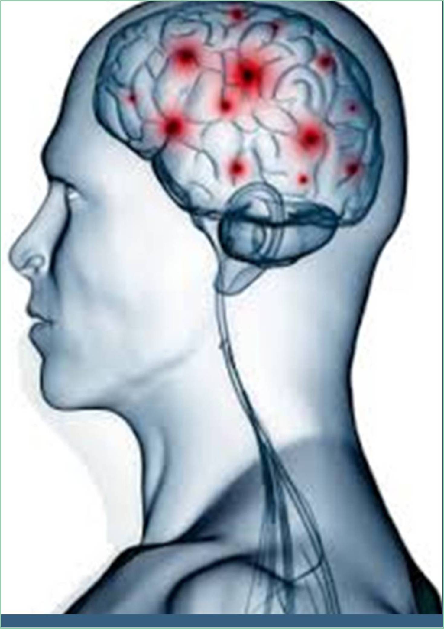
Amyloid plaque also known as neuritic plaque, Aβ plaque, or senile plaque is an extracellular deposit of the protein Amyloid beta (Aβ), which is primarily found in the grey matter of the brain. Degenerative neuronal elements and abundant microglia and astrocytes may be associated with amyloid plaques. Although some spots appear in the brain as a result of aging, numerous spots and neurofibrillary tangles are characteristic of Alzheimer’s disease. The abnormal neurites of amyloid plaques are winding, often swollen, axons and dendrites. Neurites contain a variety of organelles and debris, many of which contain the characteristic pair of spiral filaments that are the hyperfine components of neurofibrillary tangles. Plaques come in a wide variety of shapes and sizes. Aβ immunostained tissue sections consist of a lognormal size distribution curve with an average plaque area of 400-450 square micrometres (μm2). The smallest plaques (less than 200 μm2) are particularly abundant and are often composed of diffuse deposits of Aβ.
The apparent size of the plaques is influenced by the type of stain used to detect them and the plane from which they are cut for analysis under a microscope. Plaques are formed when Aβ is accidentally folded and aggregated into oligomers and longer polymers, the latter being characteristic of amyloid. Misfolded and aggregated Aβ is thought to be neurotoxic, especially in its oligomeric state. Amyloid plaques are aggregates of misfolded proteins that form in the spaces between nerve cells. These abnormally structured proteins are thought to play a central role in Alzheimer’s disease. Amyloid plaques first occur in areas of the brain that are involved in memory and other cognitive functions. Amyloid plaques are formed when fragments of a protein called beta amyloid aggregate. Amyloid beta is produced when a much larger protein called Amyloid Precursor Protein (APP) is broken down.
APP is composed of 771 amino acids and is cleaved by two enzymes to produce beta-amyloid. Large proteins are first cleaved by beta secretase and then by gamma secretase to produce beta-amyloid fragments with a length of 38, 40, or 42 amino acids. The 42-amino acid betaamyloid is chemically “sticky” than other lengths and is therefore more likely to form plaques. Studies show that each of the three genetic abnormalities associated with early-stage Alzheimer’s disease alters gamma secretase function in a way that increases beta-amyloid production 42. It is not entirely clear how beta-amyloid causes toxic damage to nerve cells, but some studies have shown that beta-amyloid can break down into fragments and release free radicals, which can attack neurons. It suggests that there is sex. Another theory is that betaamyloid forms small holes in the nerve cell membrane, causing a disordered influx of calcium that can lead to nerve cell death. Neurons die as a result, regardless of the exact pathological process by which beta-amyloid causes neuronal damage. Next, a plaque consisting of a mixture of these degenerated neurons and beta-amyloid aggregates is formed.
These plaques gradually build up in the brain because the body cannot break down and remove them. This accumulation of amyloid causes amyloidosis, which is thought to contribute to many mythological degenerative diseases. Amyloid plaques form one of the two hallmarks of Alzheimer’s disease, the other is neurofibrillary tangles. Beta amyloid is also thought to be involved in the formation of these tangles, which in turn damage neurons and cause symptoms of dementia. Technically, a person can have all the characteristics of Alzheimer’s disease, but if brain biopsy or positron emission tomography does not show the presence of amyloid plaques or neurofibrillary tangles, the diagnosis of Alzheimer’s disease is lined up.
Several therapies are currently being developed and tested to remove beta-amyloid or interfere with its production from APP. Approaches to prevent beta-amyloid production include targeting the beta-secretase and gamma-secretase needed to make it from APP. Several studies in animal models have shown that the action of these two enzymes can be successfully prevented, and some drugs based on this mechanism have reached Phase III trajectories. Another approach being studied is to prevent beta-amyloid aggregation to prevent plaque formation. Several drug candidates have been identified that appear to prevent aggregation of this protein, and these drugs are currently set to be tested in animal models of Alzheimer’s disease.